Over the past few months, the V|LF-Spiro3D project has achieved significant milestones across various Work Packages. From advancing data collection and launching new recruitments to submitting key scientific contributions, the project continues to make strides in understanding and visualizing breathing dynamics.
WP1
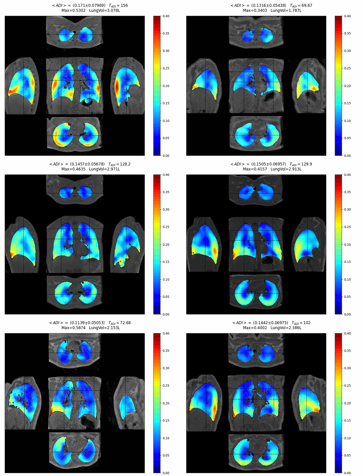
The first study, carried out at UPSaclay on 25 healthy volunteers, revealed nominal functional patterns of healthy human respiratory dynamics using 3D MR spirometry. These results formed part of Nathalie Barrau’s PhD thesis. They are about to be published in European Radiology.
3D MR spirometry was formerly implemented in every clinical site at standard magnetic field (1.5 T and 3 T), enabling the initiation of protocols for children with asthma, bronchopulmonary dysplasia or cystic fibrosis at Erasmus Medical Centre. Protocols were also initiated for adults with severe asthma at Bicêtre Hospital, chronic obstructive pulmonary disease (COPD) at Pitié-Salpêtrière Hospital and lung transplants at Foch Hospital.
Gas redistribution was revealed by 3D MR spirometry after the introduction of a biotherapy on a patient with severe asthma, supporting the clear clinical improvement experienced by the patient, while standard spirometry remained unchanged (Gharmaoui et al. SFRMBM 2025).

Tidal volume mean maps averaged over 25 healthy volunteers
WP2
The lungs are now modelled using generated 1D bronchial trees with 3D specifications for each generation, in order to simulate gas flow and distribution throughout the organ (Herszkowicz et al., SFRMBM 2025). Meanwhile, the underlying pleural pressure distribution can be computed using a statistical model.
The reconstruction of large datasets acquired by the five V|LF-Spiro3D clinical sites, covering four magnetic field strengths (0.1 T, 0.55 T, 1.5 T and 3 T) and six different MRI systems manufactured by two companies (Siemens Healthineers and GE HealthCare), remains an ongoing challenge. Production and development pipelines are being maintained and optimised at UPSaclay. Deep learning has been introduced first to speed up the process at the segmentation (Barrau et al., IABM 2024) and registration (Quique et al., IABM 2025) stages, and second, to generate reference 3D healthy lung volumes at different respiratory phases (Vaurs et al., IABM 2025, SFRMBM 2025). Alternative algorithms and approaches are still being tested, which could significantly reduce computing time.
Normal strains, i.e. how much lung tissue expands over a respiratory cycle, were tested on the datasets acquired in WP1 from patients with asthma, both before and after the administration of a bronchodilator. These strains proved to be effective markers of asthma reversibility, significantly increasing after the administration (Duwat et al., SFRMBM 2025).

Artificial 1D airway tree associated regions
WP3
Progress has been made on all aspects of WP3 in the last six months. The whole-body 0.55T scanner has been commissioned at Paris-Saclay, with sequences ported and performance characterised. The joint effort between Siemens and UPSaclay has resulted in the first dynamic lung images.
In parallel, very-low-field hardware developments are ongoing between NMR Service and AMT, where major sources of noise and artefacts have been identified and still require corrective measures. First dynamic lung images at 0.1T have also been obtained in Aberdeen, and now AMT and Paris-Saclay are advancing the post-processing pipelines to generate ventilation maps in healthy lungs.
The consortium has achieved major milestones so far: low- and very-low-field MR spirometry is no longer just a dream, and initial results are expected as planned within the next year.

Caussin et al. 4D Lung MRI across magnetic field strengths: a multicenter healthy volunteer study at 0.55T, 1.5T, and 3T accepted at ESMRMB 2025, Marseille, France
WP4
In WP4, the analysis of 10 interviews conducted in Paris is currently underway, and Irene Groenevelt and Claire Barakat are preparing a report based on the questionnaires collected. In Rotterdam, 15 participants have been involved.
POLETP is organizing a series of workshops on the a-gb-spiro3D and a-ast-spiro3D protocols, involving volunteers, patients, and medical professionals to gather experiential feedback. These workshops will be carried out at the end of November 2025. A journal article titled Imag(in)ing breathing bodies (Groenevelt et al.) is ready for submission to the Journal of Medical Humanities. As a follow-up, Irene Groenevelt plans to conduct a visual analysis focusing on color and representation in the images produced by 3/4D MR spirometry.
Remote detection of the cardiac frequency was successfully tested using spectrum analysis and peak detection through the Mediapipe platform (Figure). The outcomes critically depend on the light conditions. Work is being carried to correct for them.

WP5
On the dissemination front, the project has been showcased at numerous conferences, where preliminary findings have been shared with the scientific community. A monthly multidisciplinary seminar continues to foster collaboration and knowledge exchange across partner institutions. In terms of communication, the project’s social media presence remains strong, having reached around 20,000 people and generated over 5,000 interactions to date. This sustained engagement highlights the growing interest in and visibility of V|LF-Spiro3D, whose website has attracted more than 1500 visitors from 55 countries.


