

Oral paper presentation on MRI data postprocessing in the framework of the V|LF-Spiro 3D project. The presentation wag given at the SPIE Medical Imaging conference, in San Diego, USA, on 19 February 2024 by Catalin Fetita (IPP) and concerned lung lobes segmentation for UTE-MRI data. The title of the presentation is “Vessel-based lung lobe partitioning in ultra-short time echo proton MRI for regional ventilation assessment“.
You can read the abstract below:
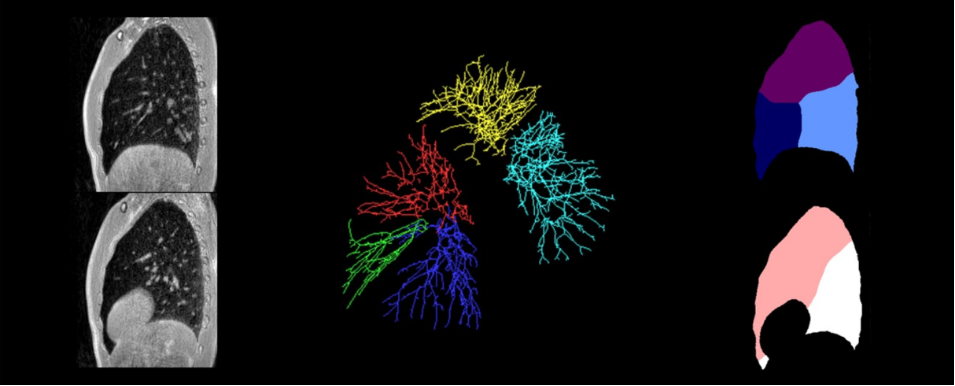
This paper addresses the problem of lung lobe partitioning in ultra-short time echo (UTE) MRI acquisitions, which are recently used for lung ventilation assessment with MRI spirometry. Because of the low image contrast, which does not enable the lung fissures display, the developed approach relies only on the vascular structures which still can be segmented from these images. The vascular network is segmented in lobes in order to generate reference clusters used for lung space partitioning. A point cloud representing the unstructured points of the vascular medial axis is partitioned in five lobes exploiting the PointNet++ framework.
The PointNet++ model is trained on data extracted from CT acquisitions and labeled using the airway and vascular trees connectivity. The airway tree lobes will define the lung lobar regions, which are propagated on the vessel structure to achieve the complete vascular labeling. A separate model is trained for the right and left lungs in order to alleviate for limited input point cloud size imposed by the model architecture and reach a high precision in classification. The trained model is applied to UTE-MRI data to generate, for a given subject, a point cloud reference that will be used for vascular lobes clustering, which will be then exploited for lung space partitioning in lobes. The approach was quantitatively evaluated on 10 CT volumes from LUNA16 dataset and qualitatively tested on additional 25 CT and 15 UTE-MRI datasets. The analysis of CT data results shows pertinent lung partitioning with respect to the lung fissures, even if a precise fissure localization is not achieved. Such result is however expected, since no information related to the lung fissure is exploited in our method because this would not be applicable to UTE-MRI data. Nevertheless, the proposed partitioning respects the vascular lobes and, to the best of our knowledge, is novel for lung MRI sequences making it possible the regional investigation of ventilation parameters in MRI spirometry.
Read the full content here!


")

")


