

During the Paris consortium meeting, groups of five to ten people were invited to participate in four workshops at BioMaps (UPSaclay) to cover 3D MR spirometry at large.

First, Adrien Duwat and Georges Willoquet presented the second prototype of the MR-compatible spirometer they are currently developing at the laboratory to allow real-time monitoring of the subject’s flow and volume at the mouth during the MR acquisition. Both electronics and interface are ready for implementation in the bore of the MR system. Participants could give it a blow and see their flow-volume loops drawn
on the screen.

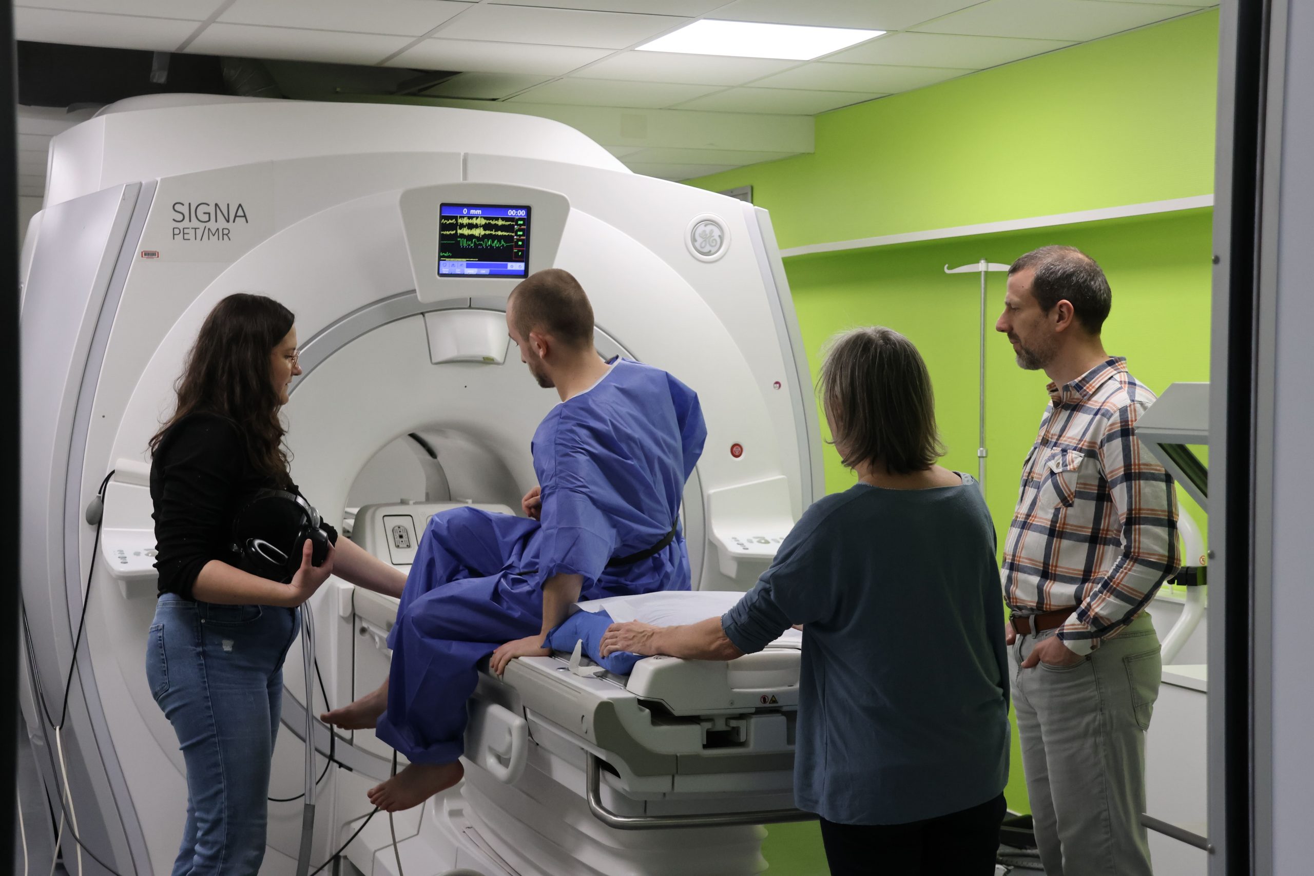
Second, Angéline Nemeth, Claire Pellot-Barakat, and Antoine Loquet performed 3D MR spirometry acquisitions on a healthy volunteer in the PET-MR GE 3 T at BioMaps: Positioning the volunteer, the respiratory belt, the thorax coil, interacting with the volunteer, starting the manual prescan and the acquisition while checking the gradient shapes on an oscilloscope, and hoping for the best out of the k-space that shows
up on the screen at the end of the scan.

Third, Ithar Gharmaoui and Anna Reitmann took every group into a short journey through the reconstruction of 3D lung dynamics over 32 retrospectively-gated respiratory phases and the subsequent data processing from deformation fields to parametric maps of flow-volume loops and associated biomarkers.

Fourth, Alexiane Pasquier and Marie Poirier-Quinot unveiled very low field approaches that are undertaken to allow large open easy access to prematured babies with a light carbon footprint with new MRI contrasts. We are talking about 10 mT, say 150 to 300 times smaller than standard clinical MRI systems. Very low field systems, which served as pedagogical tools for MRI practices, were also presented.


")

")


