Exploring the Future of Pulmonary Function Testing: Introducing 3D MR Spirometry
In the world of pulmonary testing, being able to accurately assess ventilation efficiency and diagnose respiratory diseases is really important. Traditionally, spirometry has been the go-to tool for clinicians, offering valuable insights into lung function through the measurement of forced vital capacity (FVC) and forced expiratory volume (FEV). While spirometry is easy to use and widely available, it has its limitations. One of the main problems with traditional spirometry is that it relies on the skills of the operator and the cooperation of the patient. Additionally, spirometry provides only global scalar information about lung function and is less sensitive in detecting subtle physiological impairments or assessing the extent of respiratory diseases. This can lead to inaccuracies in evaluating therapeutic efficacy, potentially resulting in misestimation of clinical benefit.

Recognising these limitations, researchers at UPSaclay have come up with a new way of testing lung function: 3D MR Spirometry. This innovative technique combines advanced imaging technology with sophisticated data analysis to provide a comprehensive assessment of how the lungs work and how they breathe in and out.
At the heart of 3D MR Spirometry is a specially designed 3D ultrashort echo time (UTE) sequence, operating at 3 Tesla magnetic field strength. This sequence captures high-resolution images with an isotropic voxel size, which allows for detailed visualization of lung structures. By acquiring images across 32 phases of the respiratory cycle and employing retrospective self-gating techniques, researchers can construct a dynamic picture of lung mechanics.

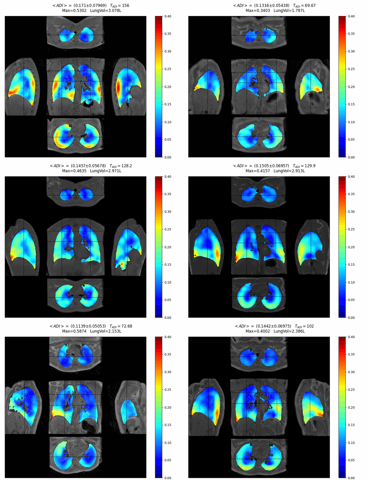
The key innovation is in the analysis of tissue displacement loops derived from the acquired images. These loops provide insights into the mechanical behaviour of the lung throughout the respiratory cycle, offering a level of detail not achievable with traditional spirometry. By computing regional flow-volume loops and mapping the Green-Lagrange strain tensor, researchers can characterise the anisotropic and hysteretic properties of lung tissue.
The advantages of 3D MR Spirometry extend beyond its ability to capture detailed physiological data. This technique isn’t limited by patient age, so it can be used on people of all ages, including children. It’s also multidimensional, which means it can extract new metrics that could be really useful for understanding respiratory diseases and assessing treatment outcomes.

In short, 3D MR Spirometry is a big step forward in pulmonary function testing. By combining cutting-edge imaging tech with top-notch data analysis, this method offers a complete picture of how the lungs work, going beyond what traditional spirometry can do. As research in this area keeps growing, 3D MR Spirometry is a great bet for improving how respiratory diseases are diagnosed and managed.


