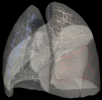
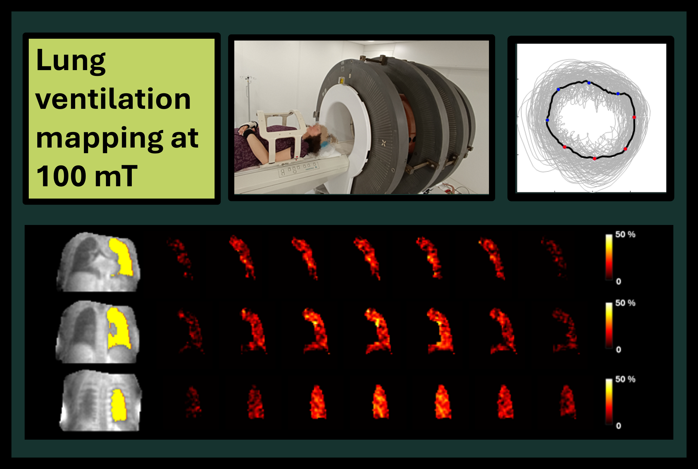

The AMT Centre has revealed its first-ever ventilation images capturing a full 3D view of the free-breathing lung using very low-field (VLF) MRI at just 100 mT. The breakthrough was presented at the ISMRM Cape Town 2026 “Take a Breath” session.
While mapping how well areas of the lung work using MRI is a promising approach for informing the treatment of respiratory diseases, access to conventional MRI scanners remains limited, restricting wider clinical use.
Taking a step towards more accessible lung imaging technologies, the AMT Centre’s latest research demonstrates the feasibility of 3D lung ventilation mapping across the full respiratory cycle at 100 mT, with a scan time of just 15 minutes. This was achieved using a novel imaging approach designed to leverage the prolonged T2* relaxation available at lower magnetic field strengths, while accommodating the hardware constraints of emerging very low-field MRI systems.
Back in Scotland, the work is already generating excitement. Dr Nicholas Senn shared the ventilation maps for the first time with the NHS Grampian respiratory disease team, where they were met with strong enthusiasm for their potential clinical impact. Building on early findings presented in 2025, Dr Senn returned to the SINAPSE Annual Scientific Meeting 2026 to showcase the full results, where he was awarded Best Session Talk.
Related ISMRM 2026 abstracts:
Senn N, et al. Pulmonary ventilation mapping using a non-commercial very-low field MRI system. Cape Town, South Africa: ISMRM 2026. no. 302-02-004 http://echo.ismrm.org/abstracts/view/739c0d55-ae32-4416-8bef-d2695f4b85ac
Senn N, et al. Free-breathing 4D lung imaging using a non-commercial very-low field MRI system. Cape Town, South Africa: ISMRM 2026. no. 560-05-006: http://echo.ismrm.org/abstracts/view/64e4754f-be0c-4fea-a398-685a88fee8fb