Functional lung imaging at 0.55 T MRI using 3D MR spirometry and Fourier decomposition to assess pulmonary complications of primary immunodeficiency diseases
50 consecutive PID patients referred to MRI of the lung for chest surveillance of airways and lung parenchyma disorders.
Objectives
Validate 3D MR spirometry against spirometry in PID patients
Assess the regional lung structural and functional changes in airways and lung parenchyma in PID patients using 3D MR spirometry combined with morphological MR images.
Compare the regional functional changes assessed using 3D MR Spirometry with regional changes in lung perfusion and ventilation obtained by Fourier decomposition.
Examination
All included patients will have standard spirometry and LF 3D MR spirometry performed the same day at Foch hospital. Standard spirometry will be performed in two positions (sitting and supine) and during two types of respiration (spontaneous and forced). Dynamic 3D lung images will be reconstructed by Siemens and processed by UPSaclay to primarily provide maps of flow-volume loops and maps of fractional anisotropy strain metric. Three to four coronal and sagittal lung perfusion- and ventilation-weighted images will be provided through Fourier decomposition.
Things happening
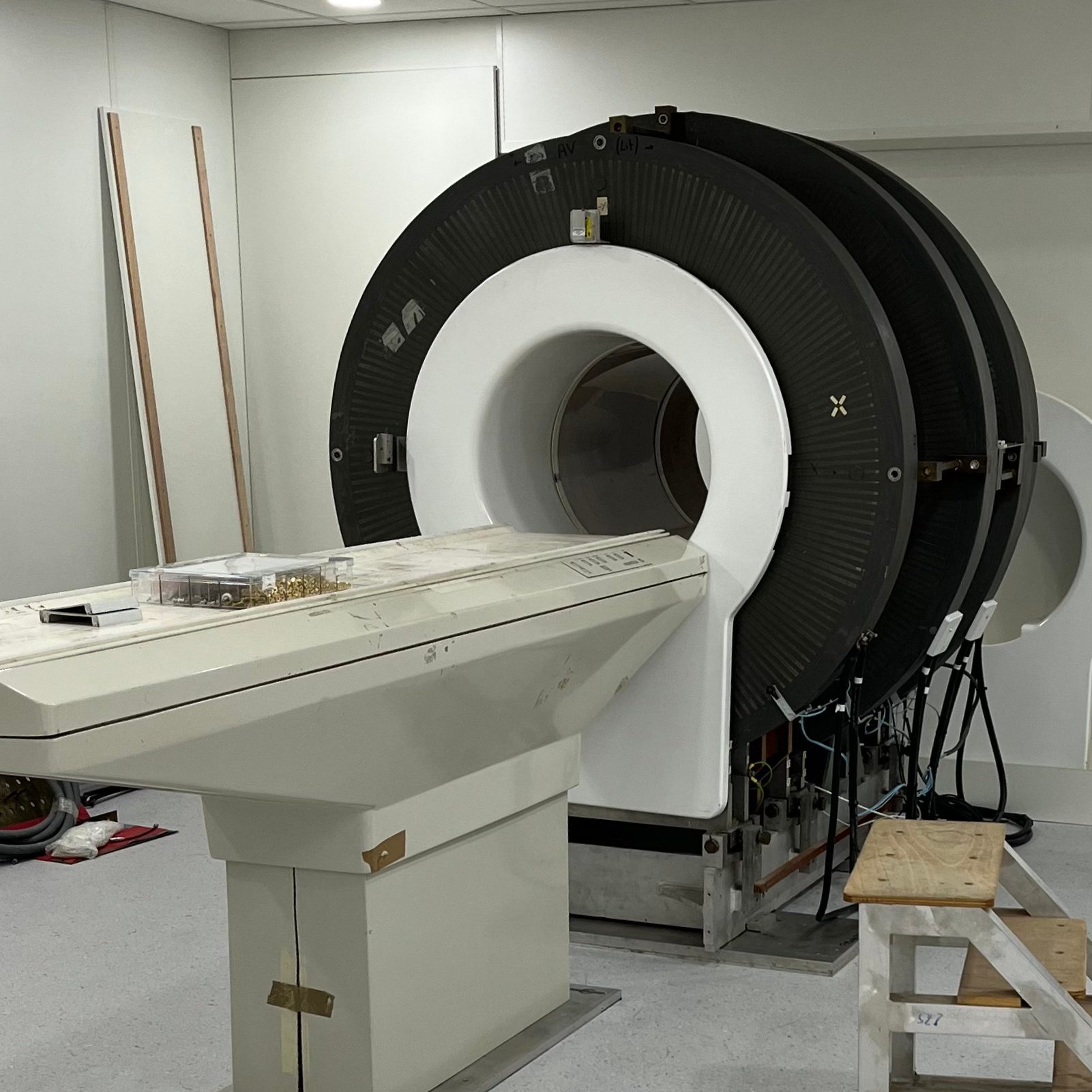
VLF whole-body scanner installation in Aberdeen
The AMT team was happy to start re-installing their very low-field MRI whole-body scanner in Aberdeen