30 healthy adults (15 women and 15 men) will be recruited to undergo LF and VLF 3D MR spirometry.
Objectives
Validate LF and VLF 3D MR spirometry against simultaneous spirometry in healthy subjects
Assess the sensitivity of LF and VLF 3D MR spirometry to prone and supine positions
Assess the regional lung structural and functional changes with prone and supine positions using LF and VLF 3D MR spirometry
Evaluate the regional lung mechanics with respect to prone and supine positions using LF and VLF 3D MR spirometry
Establish flow-volume loop maps in healthy subjects with LF and VLF 3D MR spirometry
Compare flow-volume loop maps in healthy subjects with LF and SF 3D MR spirometry
Examination
MRI and FCI scans will be acquired using a MR-compatible spirometer for reference. 3D MR spirometry will be performed on a 0.1 T MR imaging system (Magnetec) and an FCI scanner (home-made) at the Biomedical Physics Building, University of Aberdeen. We will aim to have all scans done on the same day, but to avoid strain on the volunteers the MRI and FCI examinations may be done at most within a week. Subjects will breathe freely in prone and supine positions for 2 MR acquisitions in each scanner. Dynamic 3D lung images will be reconstructed and processed (WP2) by UPSaclay to primarily provide maps of flow-volume loops and maps of fractional anisotropy strain metric.
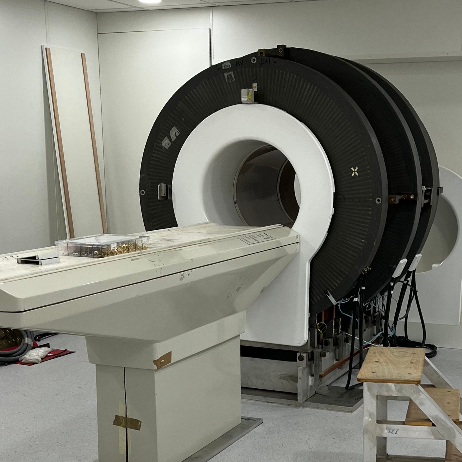
Things happening
VLF whole-body scanner installation in Aberdeen
The AMT team was happy to start re-installing their very low-field MRI whole-body scanner in Aberdeen